Overview
Acquired adult flatfoot deformity (AAFD) is a progressive flattening of the arch of the foot that occurs as the posterior tibial tendon becomes insufficient. It has many other names such posterior tibial tendon dysfunction, posterior tibial tendon insufficiency and dorsolateral peritalar subluxation. This problem may progress from early stages with pain along the posterior tibial tendon to advanced deformity and arthritis throughout the hindfoot and ankle.
Causes
The most common cause of acquired adult flatfoot is posterior tibial tendon dysfunction. What causes adult acquired flat foot? Fracture or dislocation. Tendon laceration. Tarsal Coalition. Arthritis. Neuroarthropathy. Neurological weakness.
Symptoms
The symptoms of PTTD may include pain, swelling, a flattening of the arch, and an inward rolling of the ankle. As the condition progresses, the symptoms will change. For example, when PTTD initially develops, there is pain on the inside of the foot and ankle (along the course of the tendon). In addition, the area may be red, warm, and swollen. Later, as the arch begins to flatten, there may still be pain on the inside of the foot and ankle. But at this point, the foot and toes begin to turn outward and the ankle rolls inward. As PTTD becomes more advanced, the arch flattens even more and the pain often shifts to the outside of the foot, below the ankle. The tendon has deteriorated considerably and arthritis often develops in the foot. In more severe cases, arthritis may also develop in the ankle.
Diagnosis
Looking at the patient when they stand will usually demonstrate a flatfoot deformity (marked flattening of the medial longitudinal arch). The front part of the foot (forefoot) is often splayed out to the side. This leads to the presence of a ?too many toes? sign. This sign is present when the toes can be seen from directly behind the patient. The gait is often somewhat flatfooted as the patient has the dysfunctional posterior tibial tendon can no longer stabilize the arch of the foot. The physician?s touch will often demonstrate tenderness and sometimes swelling over the inside of the ankle just below the bony prominence (the medial malleolus). There may also be pain in the outside aspect of the ankle. This pain originates from impingement or compression of two tendons between the outside ankle bone (fibula) and the heel bone (calcaneus) when the patient is standing.
Non surgical Treatment
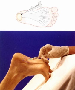
This condition may be treated with conservative methods. These can include orthotic devices, special shoes, and bracing. Physical therapy, rest, ice, and anti-inflammatory medication may be prescribed to help relieve symptoms. If the condition is very severe, surgical treatment may be needed.

Surgical Treatment
When conservative care fails to control symptoms and/or deformity, then surgery may be needed. The goal of surgical treatment is to obtain good alignment while keeping the foot and ankle as flexible as possible. The most common procedures used with this condition include arthrodesis (fusion), osteotomy (cutting out a wedge-shaped piece of bone), and lateral column lengthening. Lateral column lengthening involves the use of a bone graft at the calcaneocuboid joint. This procedure helps restore the medial longitudinal arch (arch along the inside of the foot). A torn tendon or spring ligament will be repaired or reconstructed. Other surgical options include tendon shortening or lengthening. Or the surgeon may move one or more tendons. This procedure is called a tendon transfer. Tendon transfer uses another tendon to help the posterior tibial tendon function more effectively. A tendon transfer is designed to change the force and angle of pull on the bones of the arch. It’s not clear yet from research evidence which surgical procedure works best for this condition. A combination of surgical treatments may be needed. It may depend on your age, type and severity of deformity and symptoms, and your desired level of daily activity.


 You can feel your Achilles tendon beneath the skin on the back of your ankle. It is a fibrous band of tissue that connects your calf muscles to your heel bone (calcaneus), which allows you to lift your heel off the ground. Most commonly an overuse injury, the term Achilles tendinitis commonly refers to, acute inflammation in the sheath surrounding your tendon, chronic damage to the tendon itself, called tendinosis, a combination of the two. Achilles tendinitis can range from mild inflammation to, in rare cases, a tendon rupture. One type of tendinitis, called insertional Achilles tendinitis, can affect the end of the tendon where it attaches to your heel bone. Achilles tendinitis also can be associated with other foot problems, such as painful flat feet.


You must be logged in to post a comment.